Saturday, August 27, 2016

Media Preparation and Autoclave, Plate Pouring

LAB 2 - MEDIA PREPARATION AND AUTOCLAVE, PLATE POURING


Introduction

Culture media are crucial in the controlled growth of bacteria in a laboratory. Two important requirements is that the culture media can nourish the bacteria and that it is fairly simple to make. These medias can be in the form of a liquid broth, or as a gelatinous agar. The use of each form of media will depend on whether you wish to inoculate the colony or want to visualize the bacterial colonies in a petri dish.


LB media is the most common culture media and ideal for pure conditions, made with ingredients such as tryptone, yeast, sodium chloride, and DI water. Due to the fact that LB is so rich in nutrients for bacteria, in order to prevent unwanted bacteria for eating all the nutrients, it is strongly recommended that the media is made just before autoclaving. R2A is another media that is also used for culturing bacteria, but allows the bacteria to grow slowly. R2A is the perfect media for mixed conditions, where there are slow- and fast-growing bacteria and both groups must be analyzed.

The purpose of this lab is to understand the importance of the culture media, understand and be able to operate an autoclave, and to properly and efficiently pour media into the petri dishes. Because this laboratory procedure is more oriented towards execution than analysis, there will a greater discussion on operation than on results. it is expected that some plates will not poured properly, and that some plates will be contaminated due to the amount of time they were open.


Materials and Methods

This lab was divided into tw section, with Part 1 discussing media preparation, autoclave operation and how culture medias work, while part 2 was focused on pouring premade culture media into petri dishes.
  • Part 1 was conducted verbally. The media to be used was premade. Media cultures LB and R2A were discussed, specifically the difference in nutrients that they each possess, as well as bacterial growth rate in each media. An analytical balance was used to measure 20g of LB broth, after which it was poured into a 2L flask along with 1 L of DI water. The water and brother were then mixed and the solution was distributed equally among 10 250mL beakers. Autoclave tape was then place on each beaker to indicate whether the mix was sterilized after autoclave operation or not. Autoclaving was also discussed, in terms of how it operates and how sterilization in an autoclave takes place. An autoclave was set to run for 20 minutes at 120° C. This operation can be already programmed onto the autoclave under a "LIQUIDS" setting. Water was added to the autoclave basin to create positive pressure in the chamber. Upon completion of cycle, the media was store at room temperature, while remaining closed off from the outside environment to prevent contamination.
  • Part 2 was conducted using 25 petri dishes and approximately 500mL of either LB or R2A. The culture media was poured into each dish as quickly and as effectively as possible. to provide a rough estimate, it was requested that 80% of the petri dish surface was to be covered by media and the dish be swirled around to make up for the remaining 20% of available surface area and create a thin film of culture media across the entire surface of the petri dish. A demonstration can be seen below:


Results and Discussion

To understand the importance of controlled bacterial growth in a laboratory setting, it is important to understand the vital role the culture media plays throughout the process. Important notes to be made are:
  • LB media allows for the rapid growth of microorganisms. This is due to the fact that it is extremely rich in nutrients meant to be metabolized by fast-growing bacteria. R2A is tailored to feed slower-growing microorganisms, as it uses more components and nutrients for metabolism than LB.
  • LB media is made exclusively of yeast, salt, and tryptone; R2A implements the use of yeast, peptone, dextrose, magnesium sulfate, starch, sodium pyruvate, and other components.
  • LB works really well for "pure" cultures, meaning that it is perfect for one one group of bacteria to be analyzed. R2A is better for mixed samples of bacteria, such as samples taken from the field.
  • Culture medias can be contaminated easily if left open for too long.

When it comes to the autoclave, one of the most important details is that although it is said that it is run for 20 minutes at 120­° C, it is not totally true. What really occurs is that for the sterilization to occur, the autoclave has to build temperature and pressure up until it reaches the desired temperature (120° C), and then the 20 minute timer starts. Once the timer is finished, the autoclave has to be given time to depressurize and return to room temperature. Failing to allow the autoclave to properly build pressure will not allow the culture to be sterilized, and failing to allow the autoclave to lose pressure after the sterilization will cause the positive pressure to expand rapidly, or explode and hurt the experimenter as well as the culture medias. Figure 1 below shows an autoclave with the samples in the pressure chamber.

Figure 1: Autoclave with culture media preparing for sterilization

Pouring culture media into the petri dishes is important because that is where the culture media coagulates and becomes the gelatinous film upon which the microorganisms grow. A thin film of R2A in our case was needed to cover the bottom surface of the petri dish, and it was stressed that only a little was needed to promote bacteria growth. At the same time, it was important to not get any up on the sides or lid of the dish, as it would grow bacteria on those surface as well and skew the results we would get in an actual procedure. Because pouring was so important in microbiological testing, it was important for us to practice proper pouring technique, as can be appreciated in the video above. Figure 2 shows students pouring media into the dishes, which Figure 3 shows what the final petri dish should look like with media already poured into it. Additional pictures can be appreciated below Figure 3 for documentation, observation and recording purposes.


Figure 2: (from left to right) Sebastian Arbelaez,  Alex Brawley, Jose Castano, Shane Masse, and John Price pouring media into petri dishes.


Figure 3: Final petri dish with culture media
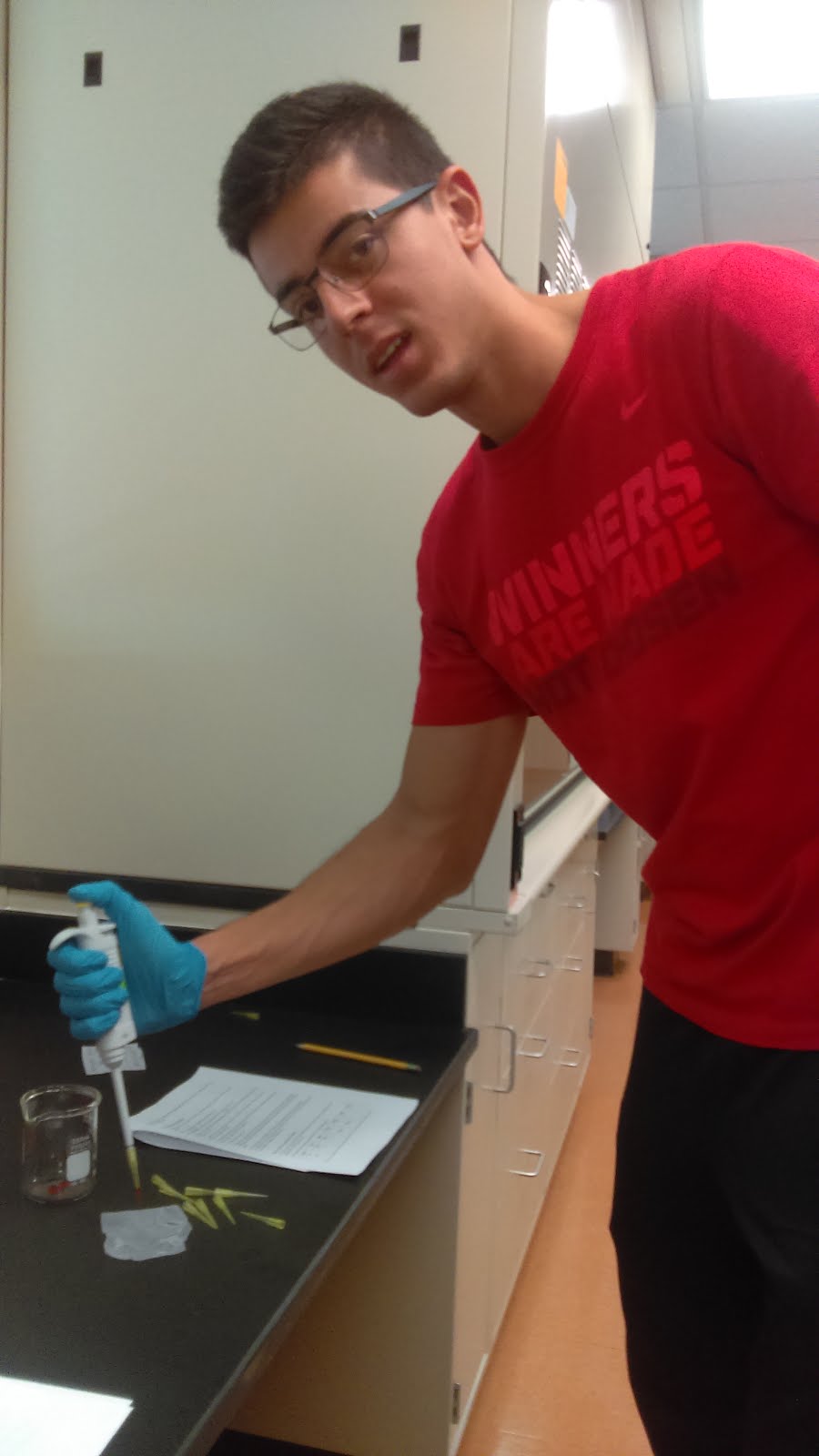
Additional figures:


Friday, August 19, 2016

Working with a Pipette

Lab 1 - Working With A Pipette


Introduction

Pipettes are essential tools used to measures specific volumes of solutions accurately and effectively. The user-friendly design of pipettes allows biologists, engineers, and chemists to easily move a solution from one vessel to another, making sure that the volume of solution needed is accurate. Pipettes are meant to be used with disposable tips that are designed for specific pipettes based on the maximum volume of solution that pipette can handle. Tips are changed in order to maintain sterility, although they can be reused if extracting from the same solution. One of the most important aspects of pipettes is making sure they are properly calibrated so that they are indeed picking up the amount of working fluid that they are supposed to.

The objectives of this lab report were to become competent in using a pipette by properly extracting/disposing solutions and to understand the importance of using an accurate pipette by analyzing the errors associated with pipettes. This laboratory procedure was divided into three parts, which allowed for learning to properly use a pipette and measuring volumes of working fluid in the correct manner. It is expected that errors will occur throughout the process, which will lead to inconsistent results and indications of improper use.


Materials and Methods
Lab 1 was performed in three sections in the following manner, with Part 1 focusing on the mechanical use of the pipette and Parts 2 and 3 focusing on the measurement of fluid using the pipette: 
  • Part 1 was conducted using a 100 μl Eppendorf micropipettor. The top knob on the pipette was rotated in order to set the volume of solution needed. A disposable tip was then attached to the end of the pipette in order to extract the solution. The plunger on the top of the pipette was pressed and held at the first stop point, as pushing it any further would contaminate the pipette and any solution to be sued after. The tip of the pipette was then inserted into the red food coloring solution, perpendicular to the surface of the solution to prevent air from forming bubbles inside the tip and reducing the amount of solution retrieved, and the plunger was subsequently released. The solution inside the pipette was then moved into a beaker, where the plunger was pushed to the first stop point to evacuate the fluid from the tip AND then to second stop point to "send a puff of air to purge the system completely of fluid". This was performed multiple times until the process for transporting solution with a pipette was understood and performed correctly.
  • Part 2 was conducted using a 100 μl Eppendorf micropipettor. A saran wrap was used as a final destination for the samples of food coloring that would be retrieved. 20 μl of red,  25 μl of yellow, 10 μl of blue, and 15 μl of green food coloring were placed on top of each other, in order to form a single drop.  The volume of the combined food coloring drops was recorded, as well as the color of the drop itself. The pipette was then reset to retrieve the recorded volume to determine whether the pipette measured the volumes correctly. 
  • Part 3 was performed using a 1000 μl micropipettor. Two test tubes were also used to contain two food coloring mixes. The first mix contained 200 μl and 300 μl of red and blue food colorings, respectively; the second test consisted of  250 μl of green and 200 μl of yellow food coloring. The final color and volume in each tube was recorded.The 1000 μl micropipettor was adjusted to the total volume in each test tube, to make sure the pipette was calibrated to the right volumes. Calibration was verified by determining if the micropipettor was able to extract all of the food coloring in each test tube.


Results and Discussion
Since Parts 2 and 3 depended on whether or not we can properly and effectively use the pipette, it was imperative that we mastered Part 1 of this lab. Many trials were performed in order to perfect the use of the pipette, which involved adjusting the angle of extraction and the use of the first and second stop positions. Figure 1 below shows the results of the three experimenters successfully using the pipette.

Figure 1. (Left to Right) Sebastian Arbelaez, Jose Castano, and Jabari Lee using a micropipettor

Part 2 heavily relied on the accuracy of the pipette to determine the amount of food coloring extracted. It was recorded that 70 μl of food coloring were used, which led to it black-colored appearance, but it was observed that the pipette left more that 25% of the food coloring still on the saran wrap, as can be appreciated in Figure 2, shown below.

Figure 2. Food coloring solution before (left) and after (right) extraction

A source of error observed by all three experimenters was that the level of fluid dropped after the plunger was completely released, indicating that air was getting into the disposable tip from the top. This means that the seal between the pipette and the disposable tip was not airtight, leading to fluid being pushed out of the tip. Other sources of error that may contribute to the inaccuracy of the pipette in retrieving the solution are:

  • the improper use of the pipette (which would include not holding the pipette perpendicular to the surface of the solution and using the plunger incorrectly),
  • an improper pipette calibration,
  • residue of solution left in the pipette tip (leading to extra solution being added to the final drop), 
  • and using the pipette too close to the saran wrap (this would cause the saran wrap to cover the tip, preventing the fluid from being extracted). 

Due to these factors of error, calibration of the pipette and the use thereof are crucial for obtaining accurate measurements during the procedure, and therefore, more accurate results in the end.


Figure 3. Food coloring solution Test Tube 1 (left) and Test Tube 2 (right).

        Part 3 followed the same practice of Part 2, with the biggest differences being the use of a larger pipette and greater volumes to be retrieved. Figure 3 depicts the final solutions in test tubes 1and 2 before the pipette was reset in order to extract shown amount of solution from each test tube. 
        It was observed that this pipette was calibrated much better than the 100 μl pipette used in Part 2; the 1000 μl pipette picked up nearly all of the fluid in both test tubes 1 and 2. It was also observed the colors within test tubes 1 and 2 were dark blue and green, respectively. Because the 1000 μl pipette performed the task of recollecting nearly all of the fluid in both cases, it was determined that the error was minimal, and that the use of the pipette was properly executed.